
The world should now look a lot less cloudy for animals that have just had cataract surgery.
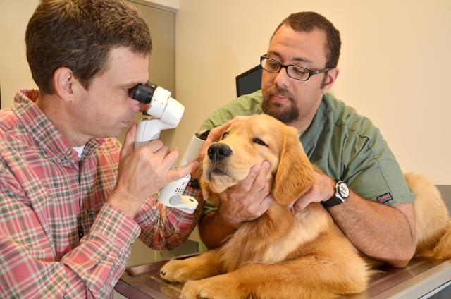
Brian Gilger , a professor of ophthalmology at the College of Veterinary Medicine, has developed a way to improve vision for both large and small animals after they have undergone cataract surgery.
When a dog has cataracts, the lens through which the eye filters light is cloudy or foggy. To fix this problem, doctors can surgically replace the faulty lens in the dog’s eyes with an artificial lens, and its vision will become relatively normal. Cataract surgery in dogs has become one of the most common and successful ocular surgeries.
The company Acrivet manufactures the artificial lenses used at the University. The lenses are made in Germany and then shipped all over the world to different veterinary facilities and ophthalmologists.
“When the lens become opaque…you take out the lens, and through a process called psycho-emulsification you can make it so the eye can focus back to the retina, and you replace that lens with an artificial lens,” Joyce Wickham, managing director and vice president of the U.S. division of Acrivet , said.
“We’ve placed intraocular lenses in animals for a long time; soluble lenses have been around for seven or eight years, and before that we used rigid lenses,” Wickham said. “With veterinary medicine, they try to adapt human ophthalmology and configure it for animals. So all the lenses are…close or very similar. [Manufacturing them] requires a lot of calculations, such as what would be the right refractions.”
Even with just an an artificial implant, multiple post-surgical complications have developed after intraocular lenses have been implanted.
Complications including ocular inflammation and development of cloudiness are often frequent symptoms after implantation. Gilger has helped to develop a method of sustained ocular release of celecoxib , an anti-inflammatory medication that can decrease ocular inflammation and eliminate these post-cataract symptoms.
In this study, special intraocular lenses containing celecoxib will be implanted in the eyes of dogs undergoing cataract surgery. The lenses, which provide a sustained release of celecoxib , were developed by the NCSU Ophthalmic Research Laboratory under Gilger’s guidance.
“The lens [in the eye] is just like [the] lens of [a] camera-it focuses the light that comes into [the] eye, but when cataracts develop, [that] is when the lens becomes opaque,” Gilger said. This is when the lens is replaced with an artificial lens, usually made of a soft-acrylic material. “The lens that is always put in after surgery… we can potentially get the lens to absorb some of this drug [ celecoxib ] and it slowly releases this drug after surgery.”
The release of the drug is a time-release formula design to prevent common post-surgery symptoms. After the entire drug has been released, the lens will continue fulfilling its purpose in the dog’s eyes for the rest of his or her life, according to Gilger.
Gilger said the modified drug-releasing lens is making its way through the necessary clinical trials to hopefully become available for everyday use in cataract surgery.
“We’ve done a number of studies evaluating the drug delivery in vitro and in models in the lab…we are inching up with a clinical trial with 20 dogs that have had cataract surgery,” Gilger said. “[In the study] some animals are receiving the drug-releasing implant and others are receiving just the implant itself, and we are studying both to see which animals are doing the best.”
If the results are positive, Gilger said they hope to use it on their clinical patients by the end of the year and then after two years hope it may be developed for commercial use. Gilger’s long-term goals are to make the product available for use in large animals and humans as well to improve their ocular health and well-being.