
Animals with missing or deformed limbs no longer face a strict sentence of amputation or disability.
Denis Marcellin-Little, professor of orthopedics at the College of Veterinary Medicine, and Ola Harrysson, professor of biomedical engineering, have teamed up to design and perform custom bone surgeries and replacements, tailored to the needs of specific dogs and cats.
“In people, there are seven sizes of total knee replacement available,” Eason Hildreth III, a 2004 graduate of CVM, said. “But there are more than seven sizes of people. Harrysson got involved in this research because his mother had a knee implant that never really worked that well for her.”
Harrysson may not be able to fix human knee implants just yet, but by perfecting a bone-modeling technique called rapid prototyping, he and Marcellin have laid the groundwork for advances in both veterinary and human medicine.
The first collaborationIt all started with Bailey, a German Shepherd born with severely deformed hind legs. She came to the Vet School for an unusual corrective bone surgery, and Marcellin wanted to design and practice the procedure before working on the actual dog.
Thanks to Harrysson and rapid prototyping, he could.
The process begins with a CT scan of the animal.
A software package called Mimics reads the CT scan as a three-dimensional image of the animal’s bones. Mimics then slices the image into a series of cross-sections guiding the construction of exact replicas of the bones, Harrysson said.
The model bones materialize from a reservoir of liquid polymer that solidifies when exposed to a laser beam. The laser sequentially traces the shape of each cross-section on the polymer. Eventually, the entire model bone solidifies, layer by layer, a millimeter at a time.
Once Bailey’s deformed bones were modeled, Marcellin could design and practice a custom surgery to correct them.
“The modeling made all the difference in the world,” Marcellin said.
Testing surgeriesIn Bailey’s case, rapid prototyping helped guide a one-of-a-kind surgery. But because bone modeling can be used to make multiple copies of the same bone, it is also a valuable research tool.
“The benefit of rapid prototyping is that we can make 27 replicates of a bone, identical in every aspect,” Hildreth said. “Then we can perform different surgical methods on the same bone and do geometric and mechanical tests to see which method was ideal.”
As a vet student, Hildreth worked with Marcellin and Harrysson to test five different surgical methods for changing the shape of a dog’s tibia, or shin bone.
The surgery, called tibial plateau leveling, restores function to dogs with injured cruciate ligaments, simply by making the destroyed ligament biomechanically unnecessary.
The tibia models used in the study were based on a Laborador Retriever named Tyler who came to the Vet School with a cruciate ligament injury.
“The middle-aged male lab is the poster child for cruciate ligament injury,” Hildreth said.
The study found two new methods for tibial plateau leveling that were equally efficient and accurate as the previously accepted technique.
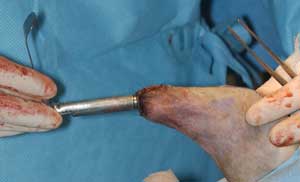
Prosthetic limbs for cats and dogsLast spring, Marcellin and Harrysson found yet another novel application for their combined expertise: installing an entire artificial limb for George Bailey, a family pet adopted as a kitten missing the lower portion of both hind legs.
Using rapid prototyping, Marcellin and Harrysson made models of George’s right leg and custom-designed a metal prosthesis that would be installed directly into the cat’s own partial leg bone.
Marcellin had to enlarge a space through the core of George’s tibia to accept a titanium nail that would anchor the prosthesis in place.
The surgery took only two hours from start to finish.
“It could have been faster,” Harrysson observed. “But there were six camera teams at the surgery and [Marcellin] had to stop at every point and let them take picutes.”
Within five days, George could step on his new leg. A few weeks later, he transformed into a rambunctious, running, jumping and chasing cat.
Unfortunately, George’s prothesis could not keep up with his enthusiastic nature. Two months after the surgery he broke thetitanium nail holding the prosthesis in place.
“If it had been a more delicate little animal, it might have been different,” Marcellin said. “But George was a little Nascar, he literally left skid marks on the kitchen floor.”
So Marcellin removed George’s broken prosthesis.
After a brief excursion into the fast lane, the cat is back where he started, no worse for the wear, and Marcellin has ideas for how to make the next prostheses last longer.
“Working with bigger ones to start with would be good,” he said. “We’ll also work with different shapes to make the bone protect the metal better.”
Making the surface of the anchor nail more porous will also encourage the bone to quickly knit directly to it, forming a lifelong interface.
Marcellin and Harrysson will soon have the opportunity to test such refinements: three dog owners have brought their pets to the Vet School for artificial legs. They are expected to undergo surgery this summer.
Human applications?Unlike George’s prosthesis, most human prostheses are now attached externally, held to soft tissue by a tight sleeve. Animal skin is more mobile, so it doesn’t offer a suitable anchor for prostheses, necessitating direct attachment to bone.
But what’s better for animals is probably also better for people.
“If you’re transferring all the load through the soft tissue, the bone starts to deteriorate,” Harrysson said.
The sleeves can also open soft tissue lesions that are hard to heal, especially in diabetic amputees.
“Soft tissue isn’t meant to take these big loads,” Marcellin said. “And bone doesn’t like not taking that load.”
So human doctors are keeping a close eye on veterinary advances in custom joint replacements and prostheses and the techniques pioneered on George may someday find their way into human hospitals.
“It’s still too risky to test in humans,” Harrysson said. “But we can fill in some of the questions now.”